Adhesive Capsulitis: The ESP Perspective on the “Frozen Shoulder”

Adhesive Capsulitis: The ESP Perspective on the “Frozen Shoulder”
At a Glance
Adhesive Capsulitis is a complex clinical syndrome characterized by the spontaneous onset of progressive shoulder pain and a significant, global loss of both active and passive glenohumeral motion. While often described as a “self-limiting” condition, the ESP approach focuses on minimizing the profound functional morbidity and psychological distress associated with its multi-year natural history.
Clinical Insight (The Sixth Sense)
The “Sixth Sense” in Adhesive Capsulitis is the ability to differentiate true capsular restriction from protective muscle guarding.
A common pitfall for generalists is over-diagnosing “Frozen Shoulder” in patients who actually have high-grade rotator cuff tears or severe subacromial impingement. The ESP distinguishes these by assessing Passive External Rotation with the patient supine and the arm at the side. If there is a firm, leathery end-feel at less than 15-20 degrees, the diagnosis is almost certainly capsular. Furthermore, “reading between the lines” of an MRI often reveals a thickened coracohumeral ligament and an obliterated axillary recess—features that confirm the diagnosis even when the clinical presentation is in the ambiguous “freezing” phase.
Anatomy & Mechanics
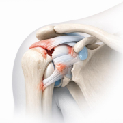
The condition primarily involves the glenohumeral joint capsule.
- The Pathological Process: Chronic inflammation leads to synovial hyperplasia and subsequent capsular fibrosis (scarring).
- The Rotator Interval: This triangular space (between the supraspinatus and subscapularis) is the epicenter of the contracture. When this area tightens, it specifically limits the “clearance” required for the humeral head to rotate, leading to the hallmark loss of external rotation.
- Biomechanical Compensation: Patients often develop “scapular hiking,” where they use the trapezius to lift the entire shoulder complex because the ball-and-socket joint itself is glued shut.
The Triage Roadmap
Red Flags (Urgent Review)
- Atypical Presentation: Pain that is worse at rest/night but not associated with movement loss (consider malignancy or Pancoast tumor).
- Systemic Symptoms: Fever, unexplained weight loss, or night sweats (potential septic arthritis or inflammatory arthropathy).
- Neurological Deficits: Significant weakness not explained by pain or stiffness.
Surgical Indicators
Surgery is rarely the first step, but the ESP-Surgeon partnership considers intervention if:
- Plateaued Progress: No measurable gain in range of motion (ROM) after 6 months of supervised conservative care.
- The “Locked” Phase: Functional impairment so severe the patient cannot perform basic activities of daily living (ADLs) or return to work.
- Procedure Options: Manipulation Under Anesthesia (MUA) or Arthroscopic Capsular Release.
Conservative Path (Evidence-Based Rehab)
The “Frozen Shoulder” requires a staged approach. Pushing through “bone-on-bone” pain in the freezing stage is counterproductive and increases inflammation.
- Phase 1 (Freezing): Pain control is priority. Gentle “pain-free” ROM. Patient education on the “thawing” timeline is the most effective intervention.
- Phase 2 (Frozen): Gradual introduction of isometric strengthening and progressive stretching within tolerance.
- Phase 3 (Thawing): High-intensity stretching and functional eccentric loading to regain end-range control.
Imaging Expectations
- X-Ray: Typically normal in Adhesive Capsulitis. Its primary purpose in the Ontario RAC pathway is to rule out glenohumeral osteoarthritis or a “locked” posterior dislocation, which can mimic the stiffness of a frozen shoulder.
- Ultrasound: May show hypervascularity in the rotator interval during the freezing phase, but is often inconclusive.
- MRI/MRA: Not routinely required for diagnosis but can show a thickened capsule (>4mm) and a lack of fluid in the axillary fold.
Interdisciplinary Outcome
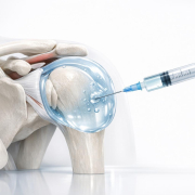
Within the ESPClinics model, the ESP acts as the primary navigator. By utilizing Image-Guided Hydrodilatation (an injection of saline and corticosteroid to physically expand the capsule) early in the “freezing” phase, we can often “short-circuit” the 18-24 month natural history. If the patient fails to respond to this interventional triad (Injection + ESP Triage + Specialized Rehab), they are fast-tracked for a surgical consultation within the Ontario Rapid Access Clinic (RAC) framework.
This model ensures that the 10% of patients who truly require a surgical release are identified early, while the 90% are managed effectively in the community without unnecessary surgical wait times.