Posterior Capsule Tightness: The Silent Driver of Impingement

Posterior Capsule Tightness: The Silent Driver of Impingement
At a Glance
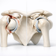
Posterior Capsule Tightness (PCT) is a common clinical finding where the fibrous tissue at the back of the shoulder joint becomes shortened or stiff. This restriction shifts the mechanics of the glenohumeral joint, often masquerading as “impingement” while being the underlying cause of persistent shoulder pain during overhead activities.
Clinical Insight (The Sixth Sense)
As an ESP, I often see patients who have been diagnosed with “Subacromial Impingement” or “Bursitis” based solely on an ultrasound report. However, the Sixth Sense of clinical reasoning tells us that the bursa isn’t the villain—it’s the victim.
When the posterior capsule is tight, it acts like a “tight hammock” that pushes the humeral head (the ball) forward and upward against the acromion (the roof) during movement. If we only treat the inflammation in the bursa without restoring internal rotation through the posterior capsule, the “impingement” will return the moment the patient resumes activity. We don’t just look at where it hurts; we look at why the space is closing.
Anatomy & Mechanics
The shoulder is a ball-and-socket joint designed for maximum mobility. The glenohumeral capsule is a redundant sleeve of tissue that stabilizes the joint.
- The Mechanism: In a healthy shoulder, as you raise your arm, the humeral head should glide inferiorly (downward).
- The Conflict: If the posterior and inferior aspects of the capsule are tight, they prevent this downward glide. Instead, the humeral head is forced superiorly, “pinching” the rotator cuff tendons and the subacromial bursa.
- GIRD: This often manifests as Glenohumeral Internal Rotation Deficit (GIRD), a hallmark sign we look for during an ESP assessment in Ontario’s Rapid Access Clinics.
The Triage Roadmap
Red Flags (Immediate Referral)
While PCT is mechanical, we must rule out “Masqueraders”:
- Night Pain: Unrelenting pain that prevents sleep regardless of position (could indicate malignancy or severe frozen shoulder).
- Fevers/Chills: Potential joint sepsis.
- Neurological Deficits: Sudden weakness or numbness in the hand/arm (Cervical Spine involvement).
Surgical Indicators
Surgery is rarely the first choice for isolated PCT. However, consultation is warranted if:
- The PCT is secondary to a Type II SLAP lesion or significant posterior labral pathology that hasn’t responded to 6 months of specialized PT.
- Mechanical “locking” or “catching” suggests an intra-articular loose body alongside the tightness.
Conservative Path (The Evidence-Based Approach)
In the Ontario ESP model, we emphasize a “Movement First” philosophy:
- Manual Therapy: ESPs or specialized PTs utilize posterior glides to “stretch” the capsule specifically.
- The Sleeper Stretch: A targeted exercise to improve internal rotation, though it must be performed with specific scapular stabilization to avoid further impingement.
- Cross-Body Adduction: Utilizing the “modified” version to isolate the capsule rather than just the deltoid.
- Kinetic Chain Integration: Strengthening the lower trapezius and serratus anterior to ensure the scapula stays out of the way of the humeral head.
Imaging Expectations
- X-ray: Usually normal, but may show a “Type II or III” acromion or minor osteophytes. It is essential to rule out glenohumeral arthritis.
- Ultrasound: Frequently shows “subacromial bursitis” or “supraspinatus tendinosis.” The ESP perspective: These are symptoms of the PCT, not the primary diagnosis.
- MRI: May show thickening of the posterior capsule or “mild posterior labral fraying.” Note that many asymptomatic people have these findings; clinical correlation is everything.
Interdisciplinary Outcome
At ESPClinics, the partnership between the Extended Scope Practitioner and the Orthopedic Surgeon ensures you don’t languish on a waitlist for a surgery you might not need.
The ESP identifies the mechanical GIRD/PCT during the initial RAC triage. If conservative management fails to restore the “Sixth Sense” of fluid motion, the ESP provides the surgeon with a detailed biomechanical map, ensuring that if an arthroscopic capsular release is required, the surgeon knows exactly which quadrant of the joint is the culprit. This streamlined Ontario pathway reduces unnecessary imaging and focuses on functional restoration.